Indocyanine green (ICG) is a green colored dye. It binds to albumin (a kind of protein), which is transported within the lymph fluid. ICG has been used to test blood flow after being injected intravenously and has also been used to show lymphatics after low dose injection to the subcutaneous tissue. ICG lymphography uses a specialist infra-red camera to detect low dose injected ICG dye in the subcutaneous tissue with the depth of 10 mm. The lymphatic function can be checked on a screen during the scan.
After ICG is injected, it will quickly be taken by the lymphatics and transported in the lymphatic tubular duct as a linear lymphatic vessel (linear fluorescence). When functioning normally, the fluid and dye will rhythmically push up the lymph proximally.
In lymphedema limb:
In lymphedema limb, the one-way perfusion may be stuck. The lymphatic fluid remains in lymphatics, and the structure of the lymphatic duct will gradually be dilated, fibrotic then obstructed. As lymphedema progresses, the fluid will leak into subcutaneous tissue, causing dermal backflow (star-like fluorescence).
Breast cancer is a very common malignant tumor that women often experience. The number of cases is increasing over the years. In addition, it can seriously threaten women’s physical and mental health. Surgery and operation are still the common treatment that doctors use. However, it can cause detrimental complications to the human body. For example, upper limb lymphedema, bring great pain to the patient and seriously affects the quality of life of the patient.
Doctor Cheng Ming-Huei, authority in plastic surgeon and ex-director of A+ Surgery Clinic, metioned that the fluorescence spectrum lymphangiography of ICG Video Scope can be used in breast cancer, breast augmentation and breast reduction. It brings applications to future clinical studies and reduces the recovery time needed after surgery. It also avoids the waste of medical resources due to the lower possibility of relapse.
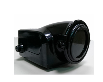
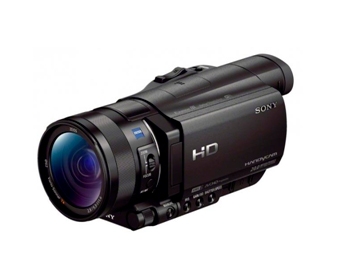
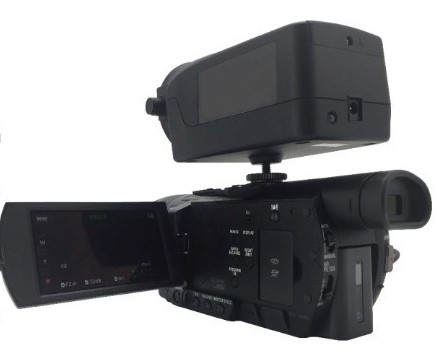
Features of ICG Video Scope
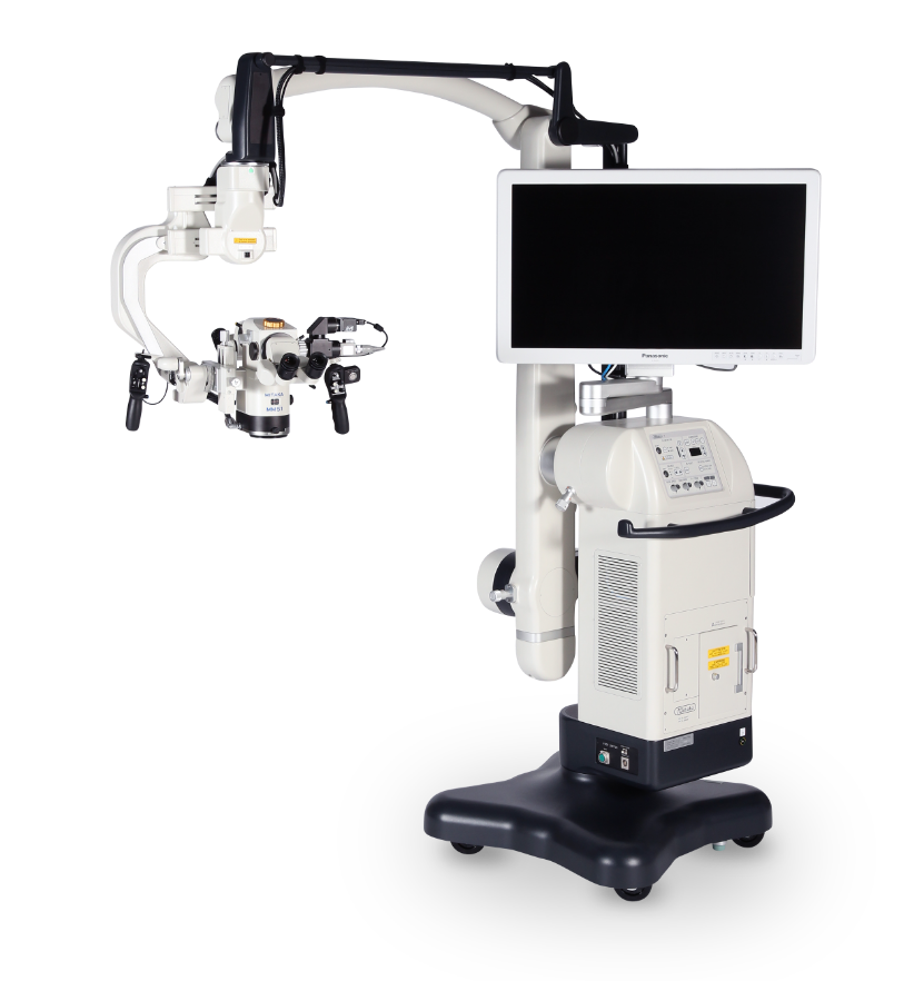
The Mitaka Surgical Microscope is high resolution at 160 line-pairs per millimeter and 42x, making it ideal for working in the sub-1mm environment.
SPY Elite, a fluorescent imaging system, may be used by surgeons to help determine whether certain tissues in the body have a strong enough blood supply for transplant purposes. Analyzing the blood circulation of tissues throughout the body may help our surgeons identify healthy donor tissue that may be harvested for such purposes, or compare the viability of various donor sites they are considering.