 1
1
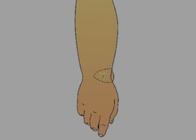
The vascularized lymph node flap is harvested from submental or groin area and transferred to the distal recipient site- dorsal wrist. The donor artery and vein are anastomosed to the recipient artery and vein under microscope. The transferred lymph node flap becomes vascularized, starts to absorb lymph from surrounding tissue, and bypasses it into the pedicle vein. As the surrounding tissue becomes dry, the catchment effect will shunt more lymph from peripheral area into the transferred lymph nodes. The gravity effect will let the lymph flow from proximal arm to the forearm naturally. The lymphedematous limb may become softer, and feel lighter from postoperative day 1. Patients do not need to wear compression garments immediately after the VLNT operation.
 2
2
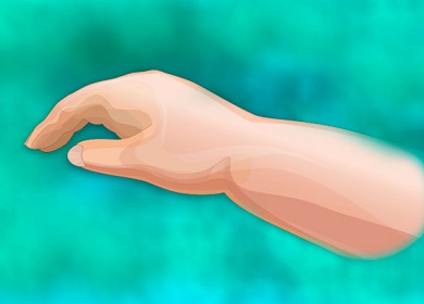
The vascularized lymph node flap is harvested from submental area and transferred to the distal recipient site- dorsal ankle. The donor artery and vein are anastomosed to the recipient artery and vein under microscope. The transferred lymph node flap becomes vascularized, starts to absorb lymph from surrounding tissue, and bypasses it into the pedicle vein. As the surrounding tissue becomes dry, the catchment effect will shunt more lymph from peripheral area into the transferred lymph nodes. The gravity effect will let the lymph flow from proximal arm to the forearm naturally. The lymphedematous limb may become softer, and feel lighter from postoperative day 1. Patients do not need to wear compression garments immediately after the VLNT operation.
 3
3
 4
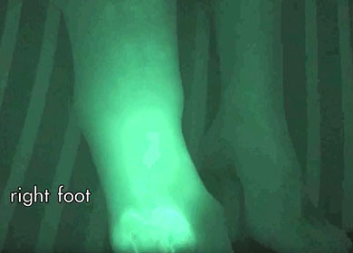
4
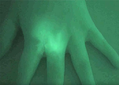
Indocyanine Green (ICG) lymphography was performed prior to surgery to detect the patent superficial lymphatic ducts. 0.5 ml 0.5% ICG is injected into both second webspacess of dorsal hands. The fluorescence is observed with the assistance of a custom-made emission device with near-infrared camcorder in 3-minutes and 20-hours. At 20-hours post- injection, the left upper limb shows diffuse dermal backflow (star dust- like fluorescence), which means the lymphatic flow of superficial lymphatic system is obstructed. A few patent lymphatic vessels (linear fluorescence) are observed in on the right upper limb, which is good for the lymphovenous anastomosis for the treatment of extremity lymphedema.
 5
5